
Fig. S3
- ID
- ZDB-FIG-170815-11
- Publication
- Garcia et al., 2017 - Sheath Cell Invasion and Trans-differentiation Repair Mechanical Damage Caused by Loss of Caveolae in the Zebrafish Notochord
- Other Figures
- All Figure Page
- Back to All Figure Page
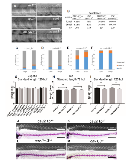
Phenotypic characterization of caveolar mutants. Related to Figure 1, 2, 3 and 4. A: DIC images of notochords of 72 hpf WT and zygotic or maternal zygotic (mz) cav1-/-, or zygotic and mz cavin1b-/-. No anatomical or notochord phenotypes were observed. Scale bars=100μm . B: Analysis of penetrance for zygotic or mz mutants. Values correspond to fraction of the fish with notochord phenotype. * Note that for the cav1,3+/- cross the penetrance at 96 hpf is higher than expected due to the fact that up to 87% of cav3-/-, cav1+/- larvae present notochord phenotype. C-F: Analysis of severity of notochord phenotype in zygotic and mz cav1, 3 and cavin1b mutants. Fish were scored as indicated (Fig 1L). n=260, 194, 82, and 279 respectively. G: Body length measurements of genotyped 120 hpf larvae from cav1,3+/- and cavin1b+/- incrosses. No significant differences were detected. One-way ANOVA, Tukey’s test, p=0.29, n=13, 54, 46, 48, 151, 117, 117, 11, 53, 47, 56, 94, 41 respectively H-I: Body length measurements of 72 and 120 hpf mz cav1, 3-/-, cavin1b-/- and cav1,3+/- and cavin1b+/-. Due to the slightly earlier onset of the notochord phenotype respect to zygotic mutants, mz mutants show a small but significant reduction in body length compared to heterozygous fish. One-way ANOVA with Tukey’s test, ***p<0.001, n=83, 81, 73, 70, 74, 81, 73, 70 respectively. J-K: Live calcein staining (top panel) and alizarin red skeletal preparations (bottom panel) of WT and mz cavin1b-/- mutant fish. No spine defects were detected (n=19 mutants). Scale bars=1mm . L-M: Live calcein staining (top panel) and alizarin red skeletal preparations (bottom panel) of WT and mz cav1, 3-/- . No spine defects were detected (n=26 mutants). Scale bars=1mm. |