
Fig. 2
- ID
- ZDB-FIG-161116-21
- Publication
- Liu et al., 2016 - A High-Content Larval Zebrafish Brain Imaging Method for Small Molecule Drug Discovery
- Other Figures
- All Figure Page
- Back to All Figure Page
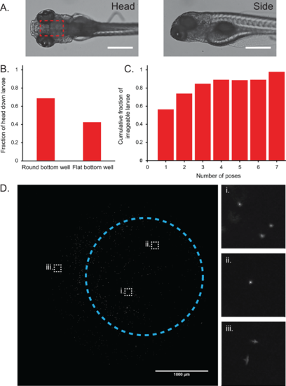
Round bottom 96-well plates preferentially orient larvae dorsal side down. (A) Zebrafish larvae must be oriented dorsal side down to image the brain region (boxed in red) using an inverted microscope. Larvae oriented side-down have their brains obscured by their large, pigmented eyes (scale bar = 500um). (B) Plots showing that round bottom 96-well plates preferentially orient fish dorsal side down for brain imaging (n = 80 larvae). (C) Approximately 90% of the larvae have at least one dorsal side-down image by the fourth reposing cycle (n = 46 larvae). (D) Maximum z-projection of 0.5um fluorescent beads used to characterize the aberrations caused by the round bottom well on an IN Cell 2000 system using a 4× 0.2 NA objective (scale bar: 1000um). Circled region indicates the typical area that a larval head rests, with the center of the circle marking the approximate center of the well. Insets show zoomed-in images of beads at different regions of the well. (i) and (ii) show no noticeable aberrations, whereas astigmatism and coma aberrations near the edges of the well are visible in (iii). |