Fig. 3
- ID
- ZDB-FIG-160414-18
- Publication
- Mishima et al., 2016 - Codon Usage and 3' UTR Length Determine Maternal mRNA Stability in Zebrafish
- Other Figures
- All Figure Page
- Back to All Figure Page
|
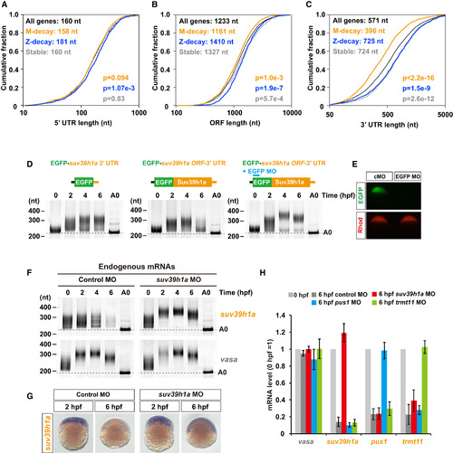
Cotranslational Deadenylation and Degradation of M-Decay mRNAs (A-C) Cumulative plots showing the distributions of 5′ UTR (A), ORF (B), and 3′ UTR (C) length in all genes (black), Z-decay genes (blue), M-decay genes (orange), and stable genes (gray). The x axis shows the length of each region on a log10 scale. The y axis shows the cumulative fraction. The median length (upper left) and the p values compared with all genes (lower right, two-sided Kolmogorov-Smirnov test) are shown. (D) Results of the time course PAT assay of EGFP reporter mRNAs injected at the one-cell stage. The developmental stages are shown above as hpf. Lanes labeled as A0 show 3′ UTR fragments without a poly(A) tail. (E) GFP fluorescence of embryos injected with GFP reporter mRNA and control MO or EGFP MO (upper panel, green). The fluorescence of coinjected rhodamine-dextran is shown as an injection control (lower panel, red). (F) Results of the time course PAT assay of suv39h1a and vasa mRNAs in embryos injected with control MO or suv39h1a MO. (G) In situ hybridization detecting suv39h1a mRNA in control MO- or suv39h1a MO-injected embryos. (H) qRT-PCR analysis of MO-injected embryos. The mRNA levels at 0 hpf were set to one. The graphs represent the averages of three independent injection experiments. The error bars show SD. See also Figures S3 and S4. |
| Genes: | |
|---|---|
| Fish: | |
| Knockdown Reagents: | |
| Anatomical Term: | |
| Stage Range: | 1-cell to Shield |
| Fish: | |
|---|---|
| Knockdown Reagents: | |
| Observed In: | |
| Stage: | Shield |

