
Fig. S10
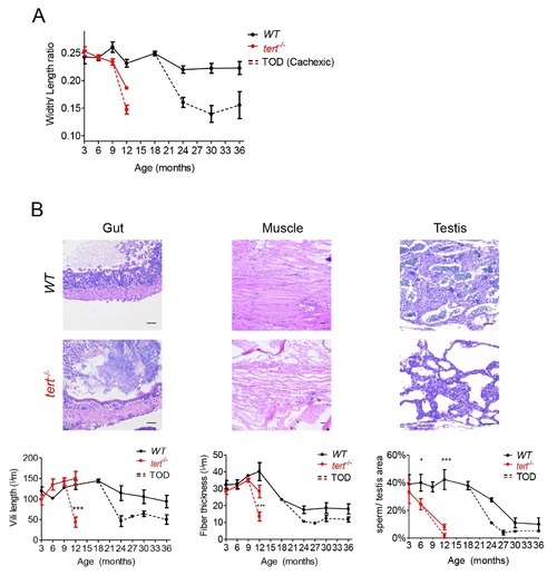
Cachexia at time of death is associated with lower body mass, gut villi shortening and marked muscle and testis atrophy.A) Quantification of width/length ratios shows that at time of death (TOD), both WT and tert-/- zebrafish have significantly lower body mass indexes when compared with non-cachexic siblings (N = 4–17 per time point for WT zebrafish and N = 3–7 for tert-/- mutants). B) Representative hematoxilin and eosin-stained sections of gut, muscle and testis from WT and tert-/- siblings at TOD. B) Cachexia is associated with shorter villi and/or villous atrophy (defined as flattening of the gut mucosal surface, N = 3–6 per genotype per time point) and severe myocyte atrophy and degeneration (N = 3–6 per genotype per time point), to a higher degree than that found for non-cachexic siblings. Testis also shows pronounced atrophy, with reduced germ cell compartment associated with cachexia (N = 3–4 per genotype per time point). TOD corresponds to the interval comprising the second and third quartiles of survival (25 to 75%). Quantifications were performed in at least 3 different fields of view for each individual. Scale bar = 50 µm. Data are represented as mean +/- SEM. |