|
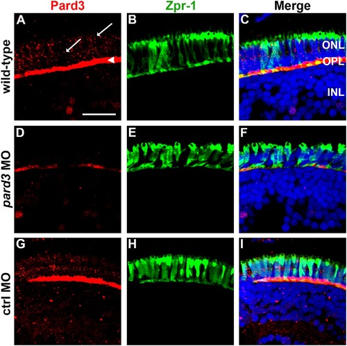
Pard3 expression is reduced at 5 dpf following injection of pard3 morpholinos. (A–C) Transverse cryosections of wild-type retinas, (D–F) pard3 morphant retinas and (G–I) control morphant retinas with Pard3 antiserum (left column) and the monoclonal antibody zpr-1 (middle column). The right panels show the merged images. Pard3 immunoreactivity was seen in the outer plexiform layer (OPL) and at cell junctions (A, arrowhead and arrows, respectively) in wild-type and control MO retinas but was significantly reduced in morphants retinas. All sections were also counterstained with DAPI (blue). ONL = outer nuclear layer; OPL = outer plexiform layer; INL = inner nuclear layer. Scale bar = 10 μm.
|

