Fig. 6
- ID
- ZDB-FIG-150526-12
- Publication
- Petersen et al., 2015 - The adhesion GPCR GPR126 has distinct, domain-dependent functions in Schwann cell development mediated by interaction with laminin-211
- Other Figures
- All Figure Page
- Back to All Figure Page
|
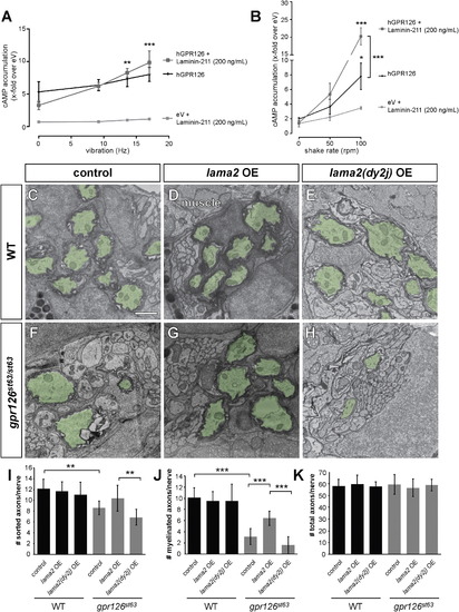
Laminin-211 Activates Gpr126 under Dynamic Conditions In Vitro and Polymerizing Conditions In Vivo (A and B) COS-7 cells were transiently transfected with empty vector (eV) and hGPR126 plasmid. cAMP accumulation was measured after stimulation with Laminin-211 (200 ng/ml) and the indicated force. Data are represented as mean ± SEM of three independent assays each performed in triplicate. p < 0.05, p < 0.01, p < 0.001, two-way ANOVA with Tukey’s multiple comparisons test (each mechanic-induced response was compared to static conditions for all hGPR126 data points, with and without Laminin-211). (A) Laminin-211 suppresses GPR126 signaling under stationary conditions (Hz 0) but causes a frequency-dependent increase of cAMP accumulation with increasing vibration. (B) Mechanical stimulation via shaking further enhances Laminin-211-dependent cAMP accumulation with increasing frequency. (C–H) TEM of 5 dpf zebrafish PLLn. Myelinated axons are pseudocolored in green. The scale bar represents 1 µm. (C–E) Myelination proceeds normally in a control larva (C), in a larva injected with 40 pg WT lama2 OE construct (D), and in a larva injected with 40 pg mutant lama2(dy2j) OE construct (E). (F) Myelination is reduced in a control-injected gpr126st63/st63 larva. (G) Injection of 40 pg lama2 OE rescues myelination in a gpr126st63/st63 larva. (H) Injection of 40 pg lama2(dy2j) OE fails to rescue myelination in a gpr126st63/st63 larva. (I–K) Quantification of TEM images. Bars represent means ± SD. p < 0.01, p < 0.001, one-way ANOVA with Bonferroni’s multiple comparisons test. (I) Number of sorted axons per PLLn. (J) Number of myelinated axons per PLLn. (K) Number of total axons per PLLn. |

