
Fig. 2, S1
- ID
- ZDB-FIG-150420-30
- Publication
- Heermann et al., 2015 - Eye morphogenesis driven by epithelial flow into the optic cup facilitated by modulation of bone morphogenetic protein
- Other Figures
- All Figure Page
- Back to All Figure Page
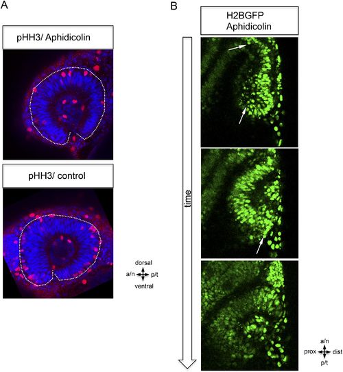
Epithelial flow is independent of cell division. (A) Retinal cell division was inhibited by application of aphidicolin, a well-established DNA polymerase inhibitor. Aphidicolin efficiently inhibited cell proliferation shown by drastically reduced pHH3 positive nuclei (upper panel, average of 6 pHH3 positive nuclei) compared to the control (lower panel, average of 21 pHH3 positive nuclei) (the optic cup is encircled with a dotted white line, 21.5 hpf). (B) We addressed the epithelial flow of aphidicolin-treated wild-type embryos, injected with H2BGFP RNA at the one cell stage; please see Figure 2A as control. The embryo was preincubated with aphidicolin 5 hr prior to the start of imaging (17 hpf, see also Video 2). The application of aphidicolin did not affect the epithelial flow. As a low level side effect of aphidicolin we observed cell death, in line with previous reports, importantly also not affecting the epithelial flow. |