
Fig. 4
- ID
- ZDB-FIG-150415-20
- Publication
- Takamiya et al., 2015 - Molecular Description of Eye Defects in the Zebrafish Pax6b Mutant, sunrise, Reveals a Pax6b-Dependent Genetic Network in the Developing Anterior Chamber
- Other Figures
- All Figure Page
- Back to All Figure Page
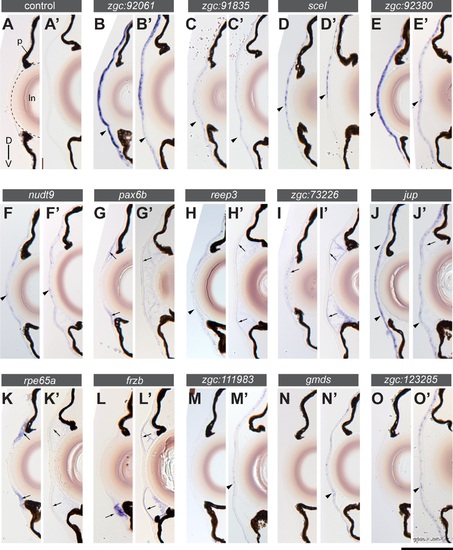
Comparison of gene expression in the cornea of wild type zebrafish at 7 dpf (A-O) and 1-month stage (A′-O′). Transverse sections with dorsal up. (A) Negative controls without probe showed no staining at both stages, except for background staining in the lens (ln, outlined by a stippled line). p: pigmentation in the iris and the retinal pigment epithelium. (B-K′) Examples of genes expressed at both 7 dpf and 1-month stage are shown. A subset of genes (B-F′) is mainly expressed in the corneal epithelium (arrowheads) and another subset (G-K′) shows expression in the corneal endothelium (arrows). (L-O′) Examples of genes that are mainly expressed at the 1-month stage. A subset of genes (M-O′) is expressed in the corneal epithelium (arrowheads) and another subset (L-L′) in the corneal endothelium (arrows). The gene symbols are indicated in the bar above each pair of sections. Scale bar: (A-O) 80 µm; (A′-O′) 100 µm. |
| Genes: | |
|---|---|
| Fish: | |
| Anatomical Terms: | |
| Stage Range: | Days 7-13 to Days 30-44 |