Fig. 3
- ID
- ZDB-FIG-140715-67
- Publication
- Sampaio et al., 2014 - Left-Right Organizer Flow Dynamics: How Much Cilia Activity Reliably Yields Laterality?
- Other Figures
- All Figure Page
- Back to All Figure Page
|
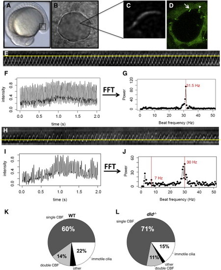
Characterization of the WT and dld/ KV Cilia Populations (A) Localization of the KV (squared region) in the body of the zebrafish embryo at 14 hpf. (B) Snapshot image of a KV of a live embryo filmed from the dorsal side. (C) Snapshot image of a KV beating cilium in a live embryo after image improved contrast for posterior kymograph analysis. (D) KV of a live embryo at 14 hpf, showing the KV cells labeled with the transgene sox17:GFP; cilia are also labeled with arl13b-GFP so that immotile cilia are bright and sharp (arrow), whereas motile cilia appear as cones with a blurred GFP label caused by the ciliary movement (arrowhead). (E) Kymograph of a cilium presenting one fundamental frequency. (F) Time series over 2 s for the same beating cilium. (G) Power spectrum analysis using FFT for the same cilium, showing that it has only one fundamental frequency of 31.5 Hz. (H) Kymograph of a cilium with two major frequencies, which we called a wobbling cilium. (I) Time series for the same beating cilium (2 s). (J) Power spectrum analysis using FFT for the wobbling cilium in (H), showing that it has two fundamental frequencies (highlighted in red): one at 7 Hz and another at 30 Hz (the lower frequency peak is an FFT artifact equal to the background noise). (K and L) Quantification of the different types of KV cilia according to motility and to CBF in (K) WT embryos, ne = 16, nc = 80, and in (L) dld-/- mutants; ne = 26, nc = 77. The class “other” includes all cilia that had a random beating pattern with no specific frequency peak after FFT analysis. KV Kupffer’s vesicle; CBF cilia beat frequency; ne number of embryos, nc number of cilia. |
| Gene: | |
|---|---|
| Fish: | |
| Anatomical Term: | |
| Stage: | 10-13 somites |
Reprinted from Developmental Cell, 29(6), Sampaio, P., Ferreira, R.R., Guerrero, A., Pintado, P., Tavares, B., Amaro, J., Smith, A.A., Montenegro-Johnson, T., Smith, D.J., Lopes, S.S., Left-Right Organizer Flow Dynamics: How Much Cilia Activity Reliably Yields Laterality?, 716-28, Copyright (2014) with permission from Elsevier. Full text @ Dev. Cell

