
Fig. S3
- ID
- ZDB-FIG-140430-27
- Publication
- Wehner et al., 2014 - Wnt/β-Catenin Signaling Defines Organizing Centers that Orchestrate Growth and Differentiation of the Regenerating Zebrafish Caudal Fin
- Other Figures
- All Figure Page
- Back to All Figure Page
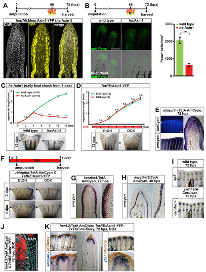
Characterization of a panel of transgenic TetON driver lines that allow for tissuespecific inducible gene expression in the adult caudal fin, related to figure 3. (A) Confocal images of YFP fluorescence in longitudinal sections of hsp70l:Mmu.Axin1-YFP (in short hs:Axin1) transgenic regenerates 6 hours post heat shock. Note that expression is ubiquitous, although levels differ greatly between cells. (B) Systemic overexpression of axin1 reduces the number of Pcna-positive cells in hs:Axin1 transgenic regenerates. A single optical section through the mesenchyme of a whole mount regenerate is shown. Error bars indicate error of the mean. 9 (C) Overexpression of axin1 starting at 3 dpa strongly interferes with regenerative growth in hs:Axin1 transgenic fish. (D) DOX treatment starting immediately after amputation does not affect regenerative growth in TetRE:Axin1-YFP transgenic fish compared to EtOH-treated controls. (E) AmCyan fluorescence and transcripts in ubiquitin:TetA AmCyan transgenic regenerates. Note that expression is fairly ubiquitous in the epidermis and the distal blastema, and covers a substantial part of the proximal blastema. (F) DOX treatment for 5 days starting 1 day before amputation interferes with regeneration in ubiquitin:TetA AmCyan; TetRE:Axin1-YFP double transgenic fish. (G) amcyan RNA expression in keratin4:TetA AmCyan transgenic regenerates is detected in the epidermis excluding the basal epidermal layer (asterisk). (H) amcyan RNA expression in keratin18:TetA AmCyan transgenic regenerates is confined to the basal epidermal layer. (I) cerulean RNA expression recapitulates endogenous sp7 expression in sp7:TetA Cerulean transgenic regenerates. (J) yfp transcripts are detected medially to the osteoblast progenitors (asterisk) in her4.3:TetA AmCyan; TetRE:Axin1-YFP double transgenic fish treated with DOX for 8 hours. Confocal image of a longitudinal sections is shown. (K) amcyan and yfp transcripts (blue) are detected proximally to mCherry (brown) in regenerates of her4.3:TetA AmCyan; TetRE:Axin1-YFP; 7xTCF:mCherry triple transgenic fish treated with DOX for 12 hours. (A-K) Small arrowheads: amputation plane. Scale bars: whole mounts, 500 μm (F) and 200 μm (B, E, I, K); sections, 100 μm. |