
Fig. 3
- ID
- ZDB-FIG-140307-54
- Publication
- Thisse et al., 1999 - Antivin, a novel and divergent member of the TGFß superfamily, negatively regulates mesoderm induction
- Other Figures
- All Figure Page
- Back to All Figure Page
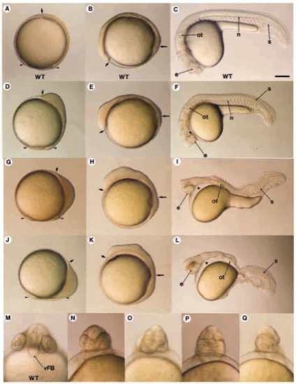
Morphological defects induced by Antivin overexpression. (A-C,M) Wild-type control (WT). (D-F) Weak phenotypes; (G-I) intermediate phenotypes; (J-L) strong phenotypes; (NQ) range of head defects. (A,D,G,J) Gastrulation stage; (B,E,H,K) somitogenesis; (C,F,I,L) 36 hours of development. (D) Embryos injected with a low dose of antivin RNA (1 pg) are characterized by a shortening and anterior thickening of the embryonic axis. (E) This thickening is located in the prospective head during somitogenesis. (F) The resulting embryos display a lack of cephalic mesoderm (star) and a range of cyclopia phenotypes as detailed in NQ. (G,H) For intermediate doses of RNA (2 pg), the embryos display a thicker and shorter embryonic axis. (I) The derived embryos lack the ventral part of the forebrain, have an unique cyclopic eye (e) and the head is devoid of all mesodermal structures (star). In the absence of notochord, somites (s) fuse under the neural tube. (J) For embryos injected with higher doses of RNA (5 pg), cells accumulate at the margin. (K) The yolk plug closure is located behind the cephalic region. (L) Embryos are defective in all ventral part of the CNS and all mesodermal derivatives of the head and trunk (star). (M-Q) Details of the deletion in the forebrain. The optical vesicles appear closer (N), fused (O-P) or a unique vesicle is formed (Q). Small arrows show the anterior tip of the embryonic axis. Long arrows, position of the yolk plug closure; arrowheads, position of the gastrula margin; n, notochord; ot, otic vesicule. Scale bar: 150 μm (A-C,G,H,L,M); 75 μm (D-F,I-K). |