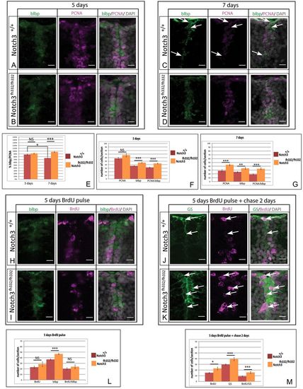
Notch3 promotes NSC quiescence and limits amplifying divisions in the juvenile pallium. (A-D) Cross-sections of the pallial ventricular zone at 5 (C,D) and 7 (A,B) dpf stained for the RG marker BLBP (green) and the proliferation marker PCNA (magenta) (gray: DAPI) in notch3fh332/fh332 homozygous mutants (B,D) and notch3+/+ siblings (A,C). White arrows at 7 dpf indicate quiescent RG. (E-G) Total number of proliferating cells (PCNA), RG (BLBP) and proliferating RG (PCNA/BLBP) per section at 7 dpf (E) and 5 dpf (F), and proportion of proliferating RG (G). (H-K) Cross-sections of the pallial ventricular zone at 5 (H,I) and 7 (J,K) dpf stained for the RG markers BLBP or GS (green) and for BrdU (magenta) (gray: DAPI) in notch3fh332/fh332 homozygous mutants (I,K) and notch3+/+ siblings (H,J). 7 dpf animals were pulsed with BrdU at 5 dpf and chased for 2 days. White arrows indicate BrdU-positive cells that remain as RG in mutants. (L,M) Total number of BrdU-positive cells (BrdU), RG (BLBP or GS) and BrdU-positive RG (BrdU/BLBP or BrdU/GS) per section at 5 dpf immediately after the BrdU pulse (L) and after 2 days of chase (M). *P<0.05; **P<0.005; ***P<0.0001 (n=3 for both notch3+/+ and notch3fh332/fh332 fish). Scale bars: 10 μm. Confocal projection images from four optical planes, each 1 μm thick.
|
