|
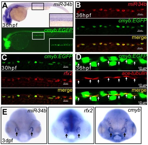
miR-34b expression is enriched in kidney MCCs and in the olfactory placode. (A) Lateral view of a 36-hpf zebrafish embryo after in situ hybridization shows that the expression of miR-34b resembles the pattern of kidney cells labeled by Tg(cmyb:EGFP). A magnified lateral view (inset) shows the labeled cells distributed in a punctate pattern. (B,C) Confocal images (lateral view) of the kidney region show the colocalization of miR-34b, rfx2 (red, WISH) and Cmyb:EGFP (green, immunostaining). (D) Confocal images of the kidney region show that each Cmyb:EGFP+ cell (green, immunostaining; overexposed to show the cilia bundle region, arrows) has a cilia bundle (immunolabeled using anti-acetylated tubulin antibody, red, arrows). The GFP staining is weaker in B and C than in D owing to the different staining methods. (E) Dorsal view of a 3-dpf embryo after in situ hybridization shows expression of miR-34b, rfx2 and cmyb in the olfactory placode (arrows). Scale bars: 20 μm in B,C; 10 μm in D.
|