|
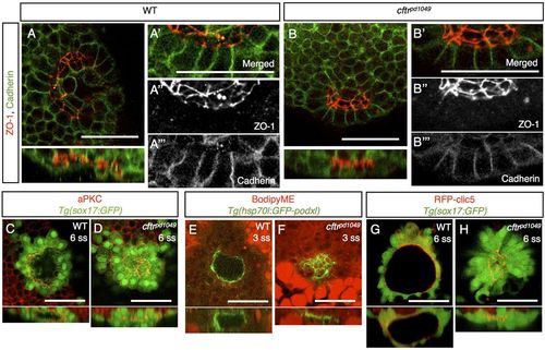
Apical-basal polarity is not affected in cftrpd1049 mutants. (A,B) Confocal image and associated orthogonal projections of WT and cftrpd1049 mutant fish stained for ZO-1 and Cadherin. (A′-A′′ ′,B′-B′′ ′) Magnification of the KV epithelium to show localization of the polarity markers. (C,D) Confocal image and associated orthogonal projections of WT and cftrpd1049 mutant fish stained for aPKC and expressing Tg(sox17:GFP). (E,F) Confocal image of WT and cftrpd1049 mutant KV expressing apically localized, GFP-tagged Podocalyxin. (G,H) Live confocal images of WT and cftrpd1049 mutant fish co-expressing an apical marker, RFP-Clic5b, and Tg(sox17:GFP). Scale bars: 50 μm.
|