
Fig. 2
- ID
- ZDB-FIG-130318-35
- Publication
- Brondolin et al., 2013 - Identification and expression analysis of the zebrafish homologs of the ceramide synthase gene family
- Other Figures
- All Figure Page
- Back to All Figure Page
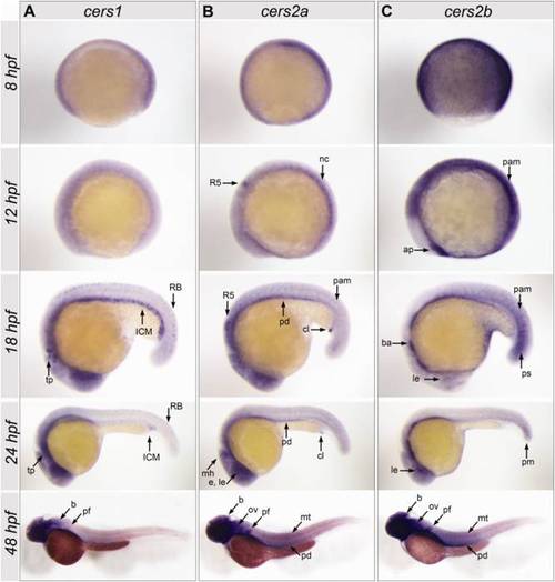
Spatial expression patterns of zebrafish cers1, cers2a and cers2b in gastrula (8 hours postfertilization [hpf]), 6-somite stage (12 hpf), 18-somite stage (18 hpf), 24 hpf, and 48 hpf embryos. A: cers1 is present in the trigeminal placode (tp), Rohon-Beard sensory neurons (RB), the hematopoietic intermediate cell mass (ICM), brain (b), and pectoral fin (pf). B: At 12 hpf, cers2a is expressed in rhombomere 5 (R5) and the developing notochord (nc), whereas at 18 hpf and 24 hpf, the gene is expressed in paraxial mesoderm (pam) and cloaca (cl), eye (e), including the lens (le), midbrain–hindbrain boundary (mh), and pronephric duct (pd). At 48 hpf, it is found in brain (b), otic vesicle (ov), pectoral fin (pf), myotomes (mt), and pronephric duct (pd). C: cers2b shows an expression in the anterior polster (ap) and paraxial mesoderm (pam), the lens of the eye (le), branchial arches (ba), and posterior somites (ps). The latter is reduced to the posterior mesoderm (pm) at 24 hpf. At 48 hpf, the expression is ubiquitous, brain (b), otic vesicle (ov), pectoral fin (pf), myotomes (mt), and the pronephric duct (pd) indicated by arrows. |