Fig. 8
|
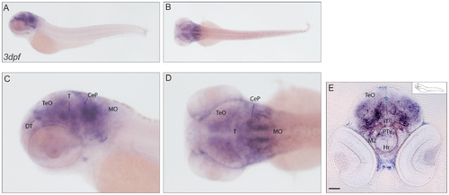
Expression pattern of cabp7a. (A–D) mRNA expression of cabp7a in lateral (A) and dorsal (B) views of 3dpf larvae with higher magnification (C–D). The signal of cabp7a antisense probe is widespread in the brain, with a stronger staining detected in the regions of the dorsal thalamus and the cerebellar plate. (E) Transverse section at the level of the eye, exhibiting diffuse expression of cabp7a in the developing brain. Scale bar: 100 µm. CeP: cerebellar plate; DT: dorsal thalamus; Hr: rostral hypothalamus; M2: migrated posterior tubercular area; MO: medulla oblongata; PTv: ventral part of posterior tuberculum; T: midbrain tegmentum; TeO: tectum opticum. |
| Gene: | |
|---|---|
| Fish: | |
| Anatomical Terms: | |
| Stage: | Protruding-mouth |

