
Fig. 2
- ID
- ZDB-FIG-130218-26
- Publication
- Janssens et al., 2013 - Matrix metalloproteinase 14 in the zebrafish: an eye on retinal and retinotectal development
- Other Figures
- All Figure Page
- Back to All Figure Page
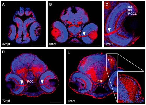
Spatiotemporal expression pattern of MMP14 protein in developing zebrafish. A-E Immunohistochemical stainings on transverse sections, made through the head of zebrafish embryos of various developmental stages show MMP14 expression (red) in the retina and the brain from 32 hpf onwards (A). At 48 hpf and 72 hpf (B-D) expression is visible in the optic nerve (white arrowhead), in the POC and in the neuropil of the OT (D, E). Higher magnification of the eye at 72hpf (C) reveals MMP14 expression in the INL, IPL and RGCL as well as in the optic nerve. The inset in E gives a detailed view of the MMP14 positive tectal neuropil. DAPI (blue) was used as counterstain. hpf, hours post fertilization; INL, inner nuclear layer; IPL, inner plexiform layer; OT, optic tectum; POC, postoptic commissure; RGCL, retinal ganglion cell layer. Scale bars: 50 μm, except inset in panel E: 100 μm. |
| Antibody: | |
|---|---|
| Fish: | |
| Anatomical Terms: | |
| Stage Range: | Prim-15 to Protruding-mouth |