Fig. 6
- ID
- ZDB-FIG-130117-10
- Publication
- Bubenshchikova et al., 2012 - Wtip and Vangl2 are required for mitotic spindle orientation and cloaca morphogenesis
- Other Figures
- All Figure Page
- Back to All Figure Page
|
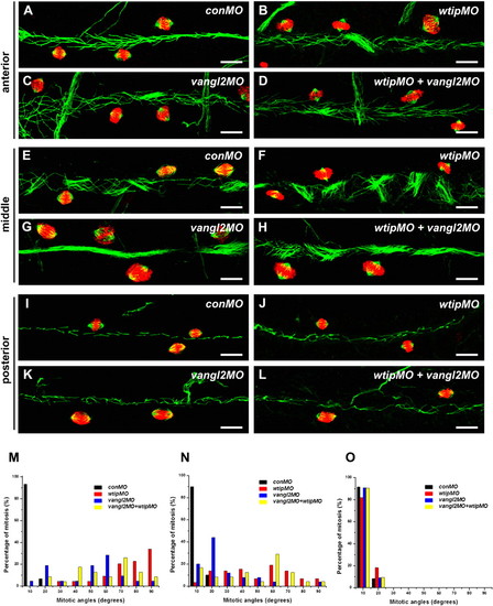
wtip knockdown leads to mitotic cell division defects in the pronephros. Whole-mount immunofluorescence of mitotic spindles for the 48 hpf pronephros were stained with anti-acetylated α-tubulin (cilia, green), anti-α-tubulin (spindle, green) and anti-phosphorylated histone H3 (pH3, red) for the anterior (A–D), middle (E–H) and posterior (I–L) pronephros. In control embryos, cells with mitotic spindle fibers oriented in the longitudinal plane of the anterior (A), middle (E) and posterior (I) pronephros. In the anterior (A–D) and middle (E–H) pronephros, wtip morphants (B,F,J), vangl2 morphants (C,G,K) and wtip + vangl2 morphants (D,H,L) resulted in mitotic spindle orientation defects. The posterior pronephros (I–K) showed mitotic spindles oriented normally in the longitudinal plan (I–L). Mitotic spindle angles were photographed by confocal imaging in the three segments of the anterior (A–D,M), middle (E–H,N) and posterior (I–L,O) pronephros at 48 hpf. The angle between the long axis of the pronephros and the spindle fibers was measured; angles were grouped into 10 degree bins. Scale bars are 100 μm in A–L. |
| Fish: | |
|---|---|
| Knockdown Reagents: | |
| Observed In: | |
| Stage: | Long-pec |

