
Fig. 2
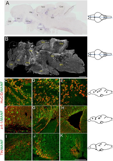
In situ hybridization mRNA analysis and immunocytochemistry of MANF expression in adult zebrafish brain. (A) A sagittal view shows manf mRNA in an adult brain section. Expression can be seen in ventral telencephalic, preoptic, ventral thalamic, pretectal, dorsal thalamic, and hypothalamic regions. (B) A sagittal view of the brain shows immuoreactivity for MANF protein. Expression is shown in telencephalic, preoptic, thalamic, optic tectal, cerebellar and rhombencephalic ventricular regions. MANF antibody co-immunostaining with HuC/D antibody in ventral telencephalic (C), dorsal thalamic (D), and rhombencephalic ventricular regions (E). Co-immunostaining with zrf-1 in dorsal telencephalon (F), ventral thalamic region (G), and telencephalic ventricle (H). Co-immunostaining with TH1 in ventral thalamic area (I), preoptic complex (J), and in caudal hypothalamus (K). MANF staining is shown in green; HuC/D, zrf-1 and TH staining are shown in red. Cce: cerebellar corpus. Ha: habenula. Hv: ventral zone of periventricular hypothalamus. OB: olfactory bulb. PPa: parvocellular preoptic nucleus, anterior part. T: thalamus. TeO: tectum opticum. TeV: tectal ventricle. Vv: ventral nucleus of ventral telencephalic area. Scale bar=300 μm in (B), 100 μm in (K). |
| Genes: | |
|---|---|
| Antibodies: | |
| Fish: | |
| Anatomical Terms: | |
| Stage: | Adult |
Reprinted from Developmental Biology, 370(2), Chen, Y.C., Sundvik, M., Rozov, S., Priyadarshini, M., and Panula, P., MANF regulates dopaminergic neuron development in larval zebrafish, 237-249, Copyright (2012) with permission from Elsevier. Full text @ Dev. Biol.