Fig. 4
|
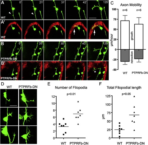
Peripheral Axons of PTPRFb-DN-Expressing RB Neurons Exhibited Axon Guidance Defects at Early Developmental Stages (A–B) Representative still images from a time-lapse confocal series showing initial peripheral axon outgrowth in WT (A) and PTPRFb-DN-expressing (B) RB neurons. The time-lapse series began at <18 SS and shows a dorsal view of the embryo. (A) and (B) show projections of complete image stacks of GFP-expressing axons; (A2) and (B2) show 5–7.5 μm optical sections of GFP-expressing axons and dsRed-expressing skin. WT peripheral axons innervated skin (A2), whereas PTPRFb-DN peripheral axons grew just below the skin (B2). Arrows indicate peripheral axon colocalization with keratinocytes. * indicates an axon branching beneath the skin. Arrowheads indicate transient filopodia from the central projection. Scale bar represents 50 μm. (C) Quantification of axon mobility during initial stages of peripheral axon outgrowth. Peripheral axons of WT and PTPRFb-DN-expressing RB neurons grew and retracted at similar rates. Error bars represent SEM. (D) Three representative peripheral axons each of WT and PTPRFb-DN-expressing RB neurons. WT axons had not yet reached the skin. Axons of PTPRFb-DN expressing RB neurons had more filopodia and did not enter the skin during the imaging period. Scale bar represents 25 μm. (E) The peripheral axons of PTPRFb-DN-expressing RB neurons had significantly (p < 0.01) more filopodia than axons of WT neurons. (F) The total filopodial length of axons in PTPRFb-DN expressing RB neurons was significantly (p < 0.05) longer than in WT axons. All data were analyzed with two-tailed t test. See also mmc4VIDEO and mmc5VIDEO. |

