
Fig. S2
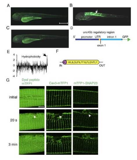
Characterization of the unc45b regulatory region. Dysferlin in silico analysis and membrane protein response to sarcolemmal damage. (A) GFP expression driven by the unc45b promoter from -1799 to -108 relative to the ATG, (B) the unc45b promoter together with the UTR, first exon and the first intron (-1799 to + 1534), (C) the unc45b intron alone upstream of the GFP coding sequence.Schematic illustration of the construct used in (B), ranging from -1799 bp to +1534 bp relative to the unc45b ATG. While the promoter and the intron alone result in weak or no muscle expression (A, C; note the expression in the yolk is unspecific), the full length construct (-1799 to +1534 bp relative to the ATG) of the unc45b gene drives robust expression in the muscle (B, arrow). The first intron of unc45b encompasses a strong transcription enhancer, resulting in specific mosaic expression in skeletal and cardiac muscle cells in transient expression assays. Orientation of embryos: anterior left, dorsal up. (E) In silico analysis of zebrafish dysferlin (Dysf) hydrophobicity (Kyte and Doolittle blot). Positive values on the vertical axis indicate hydrophobic regions. The C-terminus of the protein is towards the right and the arrow points to the transmembrane domain. The majority of Dysf is hydrophilic and the hydrophobic transmembrane domain is followed by a 22 amino acid long hydrophilic stretch at the very C-terminus. (F) Dysf transmembrane domain prediction. (G) No accumulation of Dysf C-terminal 22 amino acid peptide, caveolin 3 (Cav3-mTFP1) or mTFP1-SNAP23 (Soluble NSF Attachment Protein 23) could be observed in ruptured (arrowhead) sarcolemma. Scale bar: A-C, 500 μm; G, 4 μm. |
Reprinted from Developmental Cell, 22(3), Roostalu, U., and Strähle, U., In Vivo imaging of molecular interactions at damaged sarcolemma, 515-529, Copyright (2012) with permission from Elsevier. Full text @ Dev. Cell