|
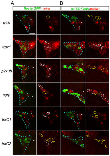
Expression of trigeminal sensory neuron markers in Trpa1b and Isl1SS subtypes. (A,B) Optical sections of 2-dpf trigeminal ganglion (outlined in blue) stained with RNA probes for sensory neuron markers (red) and antibodies against transgenic reporters (green, Trpa1b:GFP in A and Isl1SS:Kaede in B). Left panels show the merged image. Right panels show only marker staining, with transgenic reporter-positive cells outlined. White outlines indicate double-positive cells and yellow outlines mark GFP-only or Kaede-only cells. Percentages of marker-positive cells for each subtype are shown in Fig. 1E. trkC1 and trkC2 staining appears as two dots in the nucleus. Asterisk marks the adjacent lateral line ganglion. Scale bar: 20 μm.
|