
Fig. 1
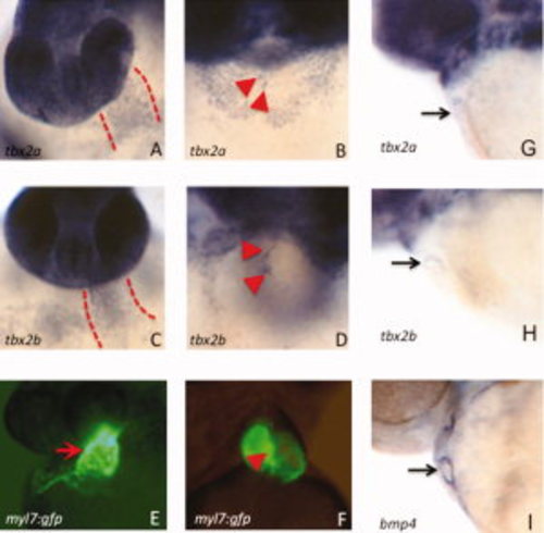
Both tbx2a and tbx2b are expressed in the primitive heart tube and transcripts become restricted to the atrioventricular canal (AVC) by 2 days postfertilization (dpf). A–D,G–I: Shown are representative embryos (n > 30 for each) analyzed by in situ hybridization to detect transcripts for tbx2a (A,B,G), tbx2b (C,D,H), or the AVC marker bmp4 (I). A–D: In addition to strong staining in the head, transcripts are readily detected in the primitive heart tube at 36 hpf (A,C, between the dashed lines) and subsequently in the AVC at 3 dpf (B,D, arrowheads marking the cushion tissue on either side of the AVC). E,F: Also shown for purpose of orientation are similarly staged myl7:gfp transgenic embryos that show the position of the heart tube at 36 hpf (E) and the AVC at 3 dpf (F). G–I: The restriction to the AVC can be seen starting already at 2 dpf, compared with bmp4. Control sense strand probes failed to generate any detectable signal under the identical conditions (not shown). |
| Genes: | |
|---|---|
| Fish: | |
| Anatomical Terms: | |
| Stage Range: | Prim-25 to Protruding-mouth |