|
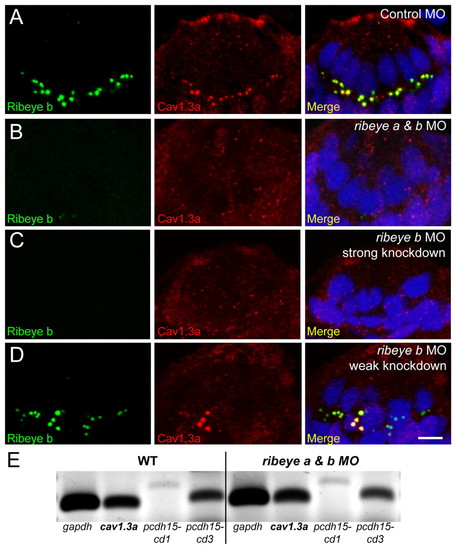
Ribeye depletion disrupts clustering of presynaptic CaV1.3a channels in hair cells. (A-D) Ribeye b (green) and CaV1.3a (red) immunolabel in cross-sections of anterior lateral line neuromasts at 4 dpf. Merged images include DAPI (blue). Scale bar: 3 μm. (A) CaV1.3a clusters are colocalized with Ribeye b-labeled punctae in control MO-injected larvae. (B) CaV1.3a clusters are absent in ribeye a & b double morphants (50 μM and 750 μM, respectively). (C) CaV1.3a clusters are absent in ribeye b MO-injected (1 mM) larvae. (D) CaV1.3a clusters in ribeye b MO-injected O (500 μM) larvae. CaV1.3a colocalized with the larger Ribeye b punctae in a neuromast with weak Ribeye b knockdown. (E) RT-PCR analysis of tails from 4 dpf larvae injected as in A and B. gapdh was used as a loading control and pcdh15 was used as a hair-cell specific transcript control.
|