
Fig. 7
- ID
- ZDB-FIG-101013-3
- Publication
- Van Otterloo et al., 2010 - Differentiation of zebrafish melanophores depends on transcription factors AP2 alpha and AP2 epsilon
- Other Figures
- All Figure Page
- Back to All Figure Page
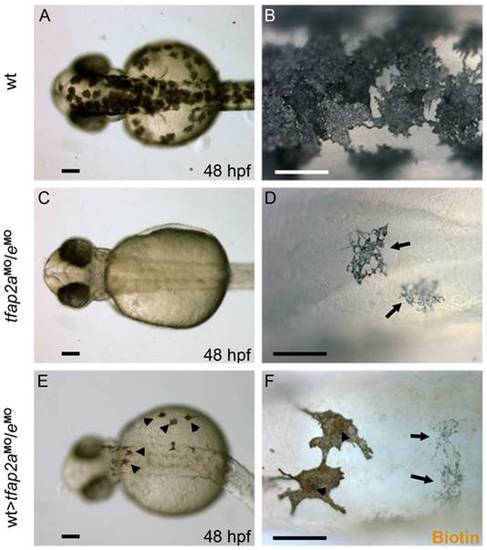
Tfap2a/e activity in melanophore differentiation appears to be cell-autonomous. (A–B) Dorsal views of a 48 hpf wild-type uninjected embryo, showing numerous, highly pigmented melanophores. (C–D) Dorsal views of a 48 hpf tfap2aMO/eMO embryo. Numbers of melanophores, and the amount of melanin per melanophore, are reduced relative to control embryos. (E–F) Dorsal views of a 48 hpf chimera generated by transplanting cells from a wild-type donor injected with biotin dextran into a tfap2aMO/eMO host, shown E) prior and F) subsequent to processing to reveal biotin. Arrowheads in E indicate normal looking melanophores. (F) Melanophores with two different morphologies are visible in this chimera. Normal-looking melanophores contain biotin (brown biotin label is most evident in the nuclei, arrowheads), indicating they are donor derived, while pale melanophores (arrows) lack biotin indicating they are host derived (In 4 embryos scored, 17 of 17 normal-looking melanophores were biotin-labeled). Scale bars: (A, C, E), 100 μm; (B, D, F), 50 μm. |
| Fish: | |
|---|---|
| Knockdown Reagents: | |
| Observed In: | |
| Stage: | Long-pec |