Fig. S5
- ID
- ZDB-FIG-101011-13
- Publication
- Garavito-Aguilar et al., 2010 - Hand2 ensures an appropriate environment for cardiac fusion by limiting Fibronectin function
- Other Figures
- All Figure Page
- Back to All Figure Page
|
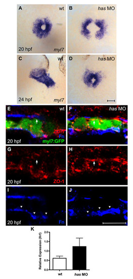
Loss of apkci function does not disrupt basal deposition of Fn. (A-D) In situ hybridization for myl7 comparing embryos injected with an anti-apkci morpholino (has MO) with their wild-type siblings. Dorsal views, anterior up. Scale bar: 50 μm. Although cardiac fusion proceeds in embryos lacking apkci function, heart tube extension is arrested (Horne-Badovinac et al., 2001; Peterson et al., 2001; Rohr et al., 2006). (E-J) Transverse confocal sections of the left lateral mesoderm in embryos expressing Tg(myl7:egfp) (green). Dorsal is up. Immunofluorescence detects ZO-1 (red) and Fn (blue). Scale bar: 10 μm. Wild-type cardiomyocytes exhibit lateral localization of ZO-1 (E,G) and basal deposition of Fn (E,I, arrowheads). By contrast, has morphant cardiomyocytes exhibit defective apicobasal polarity (Rohr et al., 2006): for example, ZO-1 is diffusely localized (F,H), including localization in the apical domain, where it is not found in wild-type cardiomyocytes (E-H, arrows). Despite the disruption of some aspects of apicobasal polarity, Fn deposition remains restricted to the basal domain of has morphant cardiomyocytes (F,J, arrowheads). Altogether, the has morphant phenotype shares many similarities with the han-/-;nat+/- phenotype. In each case, cardiac fusion proceeds, but heart tube extension arrests; additionally, the myocardium is a relatively aligned layer that exhibits basal Fn deposition despite its other apicobasal polarity defects. (K) Bar chart indicates levels of fn1 expression, relative to hprt expression, as detected by qRT-PCR in wild-type and has morphant embryos. Values represent the mean (±s.e.m.) from triplicate samples. fn1 expression is increased 2-fold in has morphants, comparable to the increase observed in han mutants (see Fig. S1C). However, our qRT-PCR data monitor fn1 expression in whole embryos, rather than in the specific locations relevant to cardiac fusion. In this regard, it is notable that the global increase in fn1 expression in has morphants does not block cardiac fusion (D) and that these embryos also exhibit normal basal deposition of Fn (J). PCR primers used were: fn1, 52-GAATCCCCGAGCCAATCAA-32 and 52-TTTTATGTTTCCCCGTTCCTT-32 hprt, 52-ATCCGCCTCAAGAGTTACCAA-32 and 52-TCTAGCAGCGTTTTCATCGTT-32. |

