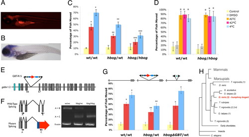
humphrey bogart (hbog) Nicotine response mutation is encoded by the zebrafish Gaba-B receptor 1.2 (gabbr1.2) locus. (A) Sagittal fluorescent imaging (anterior to left) shows a general neural expression of mRFP-gabbr1.2 fusion protein at 5 days pf. (B) Sagittal imaging (anterior to left) of a whole mount in situ hybridization for gabbr1.2 at 5 days pf (bottom) shows high levels of neural expression. (C) Full nicotine response profile of the hbog mutant comparing homozygous and heterozygous mutant animals to wild-type siblings at the standard nicotine dosage of 10 μM. Reduction from wild-type response is seen in both acute and sensitized groups. Reduction of homozygote phenotype is greater than heterozygote phenotype (P = 0.05). (D) hbog heterozygous fish show no difference in response to noxious chemicals (allyl isothiocyanate) or temperatures (4 and 42 °C) when compared to wild-type siblings. (E) Schematic representation of the GBT-R15 GBT insertion in intron 6 of the zebrafish gabbr1.2 gene. Location of exons 4 and 9 of the gabbr1.2 gene and primers A, B, and C used for RT-PCR analysis are indicated. (F) RT-PCR analysis of gabbr1.2 transcript in wild-type, heterozygous, and homozygous hbog fish. Primers in exons 4 and 9 were used to detect wild-type transcript of the gabbr1.2 gene. RT-PCR using primers in β-actin were performed as an internal control. (G) Germline propagation of Cre-mediated reversion of GBT-induced hbog mutant shows a nicotine response profile indistinguishable from wild-type sibling animals. (H) Simplified homology of the gabbr1/hbog locus shown in red. Humans and other mammals encode a single gabbr1 locus. Derived from Ensembl homology engine. *, P < 0.05 when comparing to control or acute. **, P < 0.05 when comparing to corresponding wild-type group. ***, P < 0.05 when comparing to corresponding heterozygote group and wild-type group.
|
