
Fig. 1
- ID
- ZDB-FIG-091215-7
- Publication
- Tan et al., 2009 - Regulation of membrane progestin receptors in the zebrafish ovary by gonadotropin, activin, TGF-beta and BMP-15
- Other Figures
- All Figure Page
- Back to All Figure Page
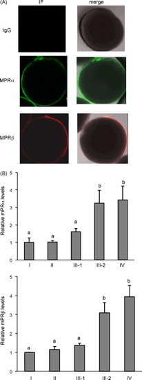
(A) Localization of zebrafish mPRα and mPRβ proteins in fully grown oocytes by immuofluorescence staining and confocal microscopy. The follicular layer was removed from zebrafish follicles and the oocytes were probed with antibodies specific for mPRα and mPRβ. Both mPRα (green) and mPRβ (red) were localized on the oocyte membrane. No immuofluorescence signal was observed using rabbit-IgG as the primary antibody. IF, immunofluorescent signals; merge, IF signal merged with a bright field picture of the oocyte. (B) Expression of mPRα and mRPβ during follicle development. Protein samples were prepared from follicles at various stages of development and subjected to Western blotting probed by mPRα, mPRβ, and α-tubulin antibodies. Data from each experiment is normalized to stage I and presented as mean ± S.E.M. (n = three experiments). Different letters denote statistical significance (p < 0.05). |
| Genes: | |
|---|---|
| Antibodies: | |
| Fish: | |
| Anatomical Term: | |
| Stage: | Days 45-89 |
Reprinted from Molecular and Cellular Endocrinology, 312(1-2), Tan, Q., Zagrodny, A., Bernaudo, S., and Peng, C., Regulation of membrane progestin receptors in the zebrafish ovary by gonadotropin, activin, TGF-beta and BMP-15, 72-79, Copyright (2009) with permission from Elsevier. Full text @ Mol. Cell. Endocrinol.