|
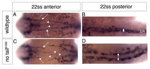
Absence of the notochord specifically affects posterior dorsal aorta development. Wildtype (A, B) or no tail b160 mutant embryos (C, D) at the 22 somite stage (ss) in situ hybridized with a probe for kdrl marking all endothelial cells. Anterior is to the left, dorsal up. A, Anterior region of a wildtype embryo showing the forming paired anterior lateral dorsal aortae (arrows), or the posterior lateral dorsal aortae (arrowheads). A, Posterior region of the same embryo as in A, showing the single posterior dorsal aorta (double arrow, DA). C, In no tail b160 mutant embryos, formation of the anterior lateral dorsal aortae occurs normally (arrows), while the posterior dorsal aortae (arrowheads indicate individual endothelial cells close to the midline) do not form properly. D, Posterior region of the same embryo as in C, showing failure of angioblasts to migrate towards the midline (double arrow, compare to double arrow in B).
|