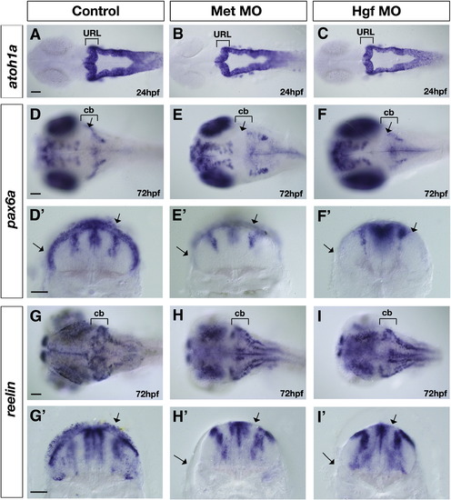
Altered expression of upper rhombic lip derivative markers in Met signaling morphants. Dorsal (A–F and J–L) and transverse views taken at the level of the cerebellum (D′–F′ and G′–I′) of control (A, D, D′, G and G′), Met morphants (B, E, E′, H and H′) and Hgf double morphants (C, F, F′, I and I′) at 24 hpf (A–C) and 72 hpf (D–I′) showing expression of atoh1a (A–C), pax6a (D–F′) and reelin (G–I′) in the cerebellum (cb), marked with brackets. The expression of atoh1a in the granule cell progenitors in the URL is not affected in Met morphants (B) and Hgf morphants (C) compared to controls (A). In contrast, the expression of markers of differentiated granule cells, pax6a (D–F′) and reelin (G–I′), is severely disrupted in Met morphants (E and E′ and H and H′, respectively) and Hgf morphants (F and F′ and I and I′, respectively) compared to controls (D and D′ and G and G′, respectively). Arrows in control embryos indicate the expression in cerebellar region (D), including expression in dorsal and ventrolateral domains in transverse sections (D′ and G′). In contrast, arrows in the Met morphants and Hgf morphants point to the absence of granule cell marker expression in both dorsal regions and in ventrolateral regions in dorsal (E and F, respectively) and transverse sections (E′ and H′ and F′ and I′, respectively). The regions of the midline at the level of the 4th ventricle and the two lateral stripes of radial glia are pax6a-positive (D′–F′) and reelin-positive (G′–I′) at 72 hpf and are not affected in Met signaling morphants (E′, F′, H′ and I′) compared to control embryos (D′ and G′). URL, upper rhombic lip; cb, cerebellum. Scale bar, 50 μm.
|
