
FIGURE
Fig. S3
- ID
- ZDB-FIG-091016-12
- Publication
- Ebarasi et al., 2009 - A reverse genetic screen in the zebrafish identifies crb2b as a regulator of the glomerular filtration barrier
- Other Figures
- All Figure Page
- Back to All Figure Page
Fig. S3
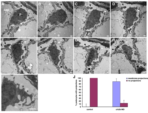
Serial reconstruction of a podocyte from a crb2b morphant. (AH) A podocyte (P) sits on top of a capillary loop (cap) and makes contact with the GBM. Apical membrane protrusions (white arrowheads) are observed which appear disorganized in structure and branch into the urinary space. When this protrusion makes contact (black arrowhead) with adjacent GBM, it appears to form foot process like structures. Ultrathin sections taken every 70 nm. (I) Higher magnification of panel F. (J) Quantification of apical projection defects in crb2b MO and controls. Scale bars, 500 nm. |
Expression Data
Expression Detail
Antibody Labeling
Phenotype Data
Phenotype Detail
Acknowledgments
This image is the copyrighted work of the attributed author or publisher, and
ZFIN has permission only to display this image to its users.
Additional permissions should be obtained from the applicable author or publisher of the image.
Reprinted from Developmental Biology, 334(1), Ebarasi, L., He, L., Hultenby, K., Takemoto, M., Betsholtz, C., Tryggvason, K., and Majumdar, A., A reverse genetic screen in the zebrafish identifies crb2b as a regulator of the glomerular filtration barrier, 1-9, Copyright (2009) with permission from Elsevier. Full text @ Dev. Biol.