
Fig. 5
- ID
- ZDB-FIG-090710-7
- Publication
- Roberts et al., 2009 - Apical polarity protein PrkCi is necessary for maintenance of spinal cord precursors in zebrafish
- Other Figures
- All Figure Page
- Back to All Figure Page
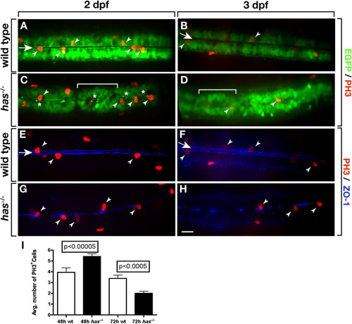
Loss of PrkCi function causes a transient excess and then deficit of dividing spinal cord precursors. A-D: Images, from dorsal view and focused on the trunk spinal cord, of wild-type and has-/- embryos and larvae carrying the Tg(olig2:egfp) reporter and labeled with anti-PH3 antibody to mark M phase cells. In wild-type most mitotic cells (arrowheads) are adjacent to the central canal (arrow). A,B: More cells divide at 2 days postfertilization (dpf; A) than at 3 dpf (B). C,D: In has-/- embryos, many EGFP+ cells form rosettes (brackets) surrounding discontinuous portions of central canal (asterisks). PH3+ are usually only found within rosettes. E-H: Images, from dorsal view and focused on the trunk spinal cord, of wild-type and has-/- embryos and larvae labeled with anti-PH3 antibody (red) and anti-ZO-1 antibody (blue) to mark apical membrane. E,F: In wild-type, ZO-1 localization outlines a continuous central canal closely associated with PH3+ mitotic cells. G,H: In has-/- embryos and larvae, ZO-1 labeling is discontinuous but mitotic cells are nearly always associated with remaining apical membrane. I: Quantification of PH3+ cells at 2 and 3 dpf over a 288 μm length of trunk spinal cord. Error bars represent SEM. Statistical significance was determined using Student′s t-test. |