
Fig. S1
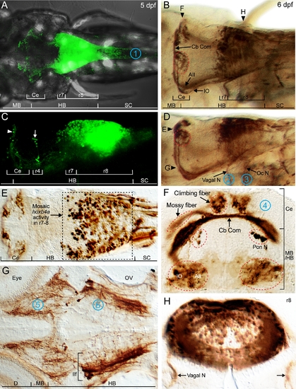
Live imaging and immunohistochemically detected hoxb4a activity in the midbrain, cerebellum, hindbrain and spinal cord. Composite dorsal (A) and side (C) views of hoxb4a expression in a live 5 dpf transgenic zebrafish from 210 μm and 150 μm confocal stacks, respectively. (B, D) Dorsal (B) and side (D) views of hoxb4a-YFP using immunohistochemistry (anti-YFP) in a fixed 6 dpf fish. Horizontal (E, G) and coronal sections (F, H) with section planes indicated in (B, D). Target sites for retrograde labeling are marked by 1 (spinal cord), 2 (Xth nerve), 3 (pectoral fin), 4 (cerebellum), 5 (midbrain) and 6 (r4). Abbreviations: AII, Area II; Ce, cerebellum; D, diencephalon; HB, hindbrain; llf, lateral longitudinal fascicle; IO, inferior olive; MB, midbrain; mlf, medial longitudinal fascicle; Oc N, occipital nerve; OV, otic vesicle; Pon N, pontine nucleus; SC, spinal cord; Vagal N, vagal nerve. B, D and E–H are cropped high magnification illustrations of Figs. 5K, 5J, 5C, 5T, 5D and 4B, respectively, (from [25]). |