
FIGURE
Fig. 8
Fig. 8
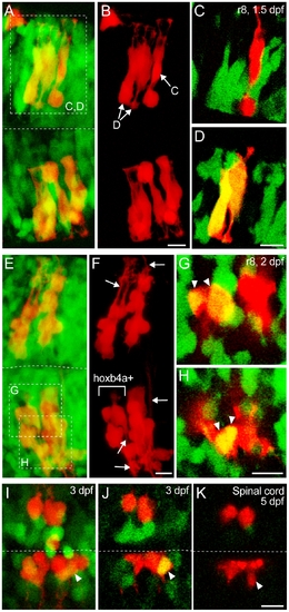
Clonal analysis by single cell injection. (A–B, E–F, I) Composite dorsal views from 165 μm (A–B), 170 μm (E–F) and 130 μm (I) confocal stacks showing progenitors from a single cell injected during the gastrula period (red) and hoxb4a-YFP (green). (C–D, G–H, J–K) Single plane high magnification images showing the progenitor cells and hoxb4a-YFP expression. Arrowheads point to the co-labeled cells. Arrows in F mark the processes (dendrite/axon) extending from the neurons at 2 dpf. Anterior is to the left. Dashed lines marked the midline. Scale bars = 10 μm. |
Expression Data
Expression Detail
Antibody Labeling
Phenotype Data
Phenotype Detail
Acknowledgments
This image is the copyrighted work of the attributed author or publisher, and
ZFIN has permission only to display this image to its users.
Additional permissions should be obtained from the applicable author or publisher of the image.
Full text @ PLoS One