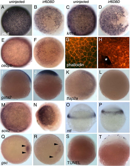
Irf6DBD-mediated changes in gene expression in shield-stage D. rerio embryos. Dorsal views, except as indicated, of uninjected and irf6DBD-injected embryos, as indicated by the column header, fixed when uninjected sibling embryos reached shield stage. Embryos processed for in situ hybridization to reveal expression of the indicated gene, except as noted. Images portray the phenotype displayed by 60%–80% of embryos in the experimental group, n = 25 embryos or greater. (A–F) Expression of zfk8, krt18, and cebpb, are expressed in all EVL cells of uninjected embryos but absent in large patches in uninjected embryos. (G) Uninjected and, (H) irf6DBD-injected embryos stained with phalloidin, revealing polymerized actin (i.e., F-actin). (G) In the uninjected embryo, F-actin is visible in a cortical ring within every EVL cell. (H) In the irf6DBD-injected embryo, there is loss (*) or disorganization of F-actin in superficial cells. (I, J) Lateral views, showing gata2 expression in non-neural epiblast is present but displaced the irf6DBD-injected embryo. (K, L) In contrast to gata2, expression of tfap2a, a marker of non-neural epiblast, is highly reduced in the irf6DBD-injected embryo. (M,N) sox2 expression, a marker of neural epiblast, is present in the irf6DBD-injected embryo. (O, P) Lateral views, showing that ntl expression, a marker of hypoblast, is present in the irf6DBD-injected embryo. (Q, R) gsc expression is present in the shield hypoblast of the uninjected embryo, and in an expanded arc in the irf6DBD-injected embryo, consistent with developmental delay in the latter (arrowheads show extent of gsc expression arc). (S, T) Lateral views of embryos processed to reveal cells undergoing apoptosis by TUNEL. TUNEL-positive cells are absent from this uninjected embryo. In the irf6DBD-injected embryo, many TUNEL-positive cells are visible, primarily within the EVL.
|
