
Fig. 5
- ID
- ZDB-FIG-081222-16
- Publication
- Kagemann et al., 2008 - Repeated, noninvasive, high resolution spectral domain optical coherence tomography imaging of zebrafish embryos
- Other Figures
- All Figure Page
- Back to All Figure Page
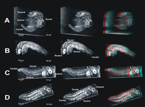
Stereo pairs of images reveal internal structures in whole embryos in three dimensions. These panels provide visualization of three dimensional (3D) data in a printed image. Such data are best visualized interactively, rotating the projected data manually to obtain an optimal view of the structure of interest; impossible in printed image (see also Figure 2). Panel A contains a 3D crossed-stereo image of a 24 hpf embryo. To view, gently cross your eyes until 3 images appear, and focus on the image in the middle. The image on the far right of panel A is the same image, and can be viewed with red/blue 3D glasses to visualize the embryo in 3D. Panel B contains a crossed-stereo pair and red/blue stereo image of a 24 hpf embryo. Panel C contains a 72 hpf stereo-pair and red/blue 3D image. In panels A-C, the embryo appears to be facing out of the image. Panel D contains a stereo-pair and red/blue 3D image of a 120 hpf embryo facing into the image. |