
FIGURE
Fig. 1
- ID
- ZDB-FIG-081222-12
- Publication
- Kagemann et al., 2008 - Repeated, noninvasive, high resolution spectral domain optical coherence tomography imaging of zebrafish embryos
- Other Figures
- All Figure Page
- Back to All Figure Page
Fig. 1
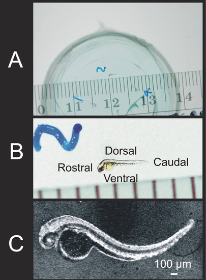
The appearance of the 72 hpf zebrafish with millimeter ruler, at magnification, and as observed by SD-OCT provided for appreciation of its small size. Zebrafish embryos were embedded in 1% agarose gel in an inverted microscopy Petri dish (A, B; B shows a magnified region of A). Embryos were scanned in three dimensions (3D), and reflectance of internal structures quantified. C-mode sections of the 3D data set could be isolated and tissue reflectance within a slice is displayed (C). |
Expression Data
Expression Detail
Antibody Labeling
Phenotype Data
Phenotype Detail
Acknowledgments
This image is the copyrighted work of the attributed author or publisher, and
ZFIN has permission only to display this image to its users.
Additional permissions should be obtained from the applicable author or publisher of the image.