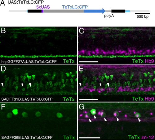
Expression of TeTxLC:CFP in double transgenic embryos. (A) The UAS:TeTxLC:CFP fish carries a single-copy insertion of T2SUASTeTxLCCFP within the CSPP1 gene. Blue boxes indicate exons. (B-G) Lateral views of the trunk of double transgenic embryos immunostained with the anti-GFP antibody (green) and the anti-Hb9 (B-E) or the zn-12 (F and G) antibody (red). Arrowheads indicate costaining with anti-GFP and anti-Hb9 (E) or anti-GFP and zn-12 (G). Anterior is to the left, and dorsal is to the top. (Scale bars: 50 µm.) (B and C) The hspGGFF27A;UAS:TeTxLC:CFP embryo at 48 hpf. The anti-GFP antibody detects the TeTxLC:CFP fusion protein but does not detect the GGFF protein in this condition (data not shown). Axons of descending hindbrain interneurons were strongly stained (green). The anti-Hb9 antibody detected spinal motor neurons (red). (D and E) The SAGFF31B;UAS:TeTxLC:CFP embryo at 48 hpf. The anti-GFP antibody detected spinal interneurons and motor neurons (green). (F and G) The SAGFF36B;UAS:TeTxLC:CFP embryo at 30 hpf. Both the anti-GFP and zn-12 antibodies detected Rohon-Beard neurons (green and red).
|
