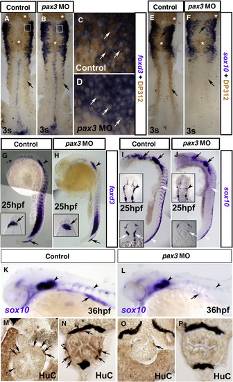
Pax3 is required for enteric nervous system development. In situ mRNA hybridisation analysis of foxd3 (A–D, G, H) and sox10 (E, F, I–L) and immunodetection of DP312 (A–F) and HuC (M–P). Dorsal flat mounts (A–F, I, J upper insets) are anterior to top. Lateral view whole mounts are anterior to top and dorsal to right (G–J) or anterior to left dorsal to top (K, L). Transverse cryosections (G–J lower insets, M–P) are dorsal to top. (A–F) Injection of pax3 MO depletes nuclear Pax3/7 immunoreaction at 3 s (B, D, F; arrows) in regions of pax3 mRNA expression, but leaves residual signal in regions of expression of pax3b, pax7 or Rx genes in the brain and eye (asterisks). Foxd3 expression in NC, tailbud and floorplate is unaffected by Pax3 knockdown (A, B; cranial NC, boxes enlarged in panels C, D). Pax3 knockdown has no effect on sox10 expression in cranial NC and otic placode at this stage (E, F). (G–J) At 25 hpf, foxd3 expression is restricted in NC to a cluster of nascent pre-migratory cells in the tail (G, H, arrows) and to lateral line primordium (G, H, arrowheads). Pax3 MO injection has no effect on foxd3 expression in either location (H), but reduces sox10 mRNA in pre–migratory crest and cells migrating on the medial pathway (I, J, white arrows), while having little effect on expression in the head (I, J, black arrows). Significant sox10 mRNA in nascent crest (I, J, white arrowheads) is down-regulated anteriorly. (K, L) At 36 hpf, sox10 mRNA is reduced in pax3 morphants in the ventral head region destined to form enteric neurons (arrows) but is still detected in otic placode and glia (arrowheads). (M–P) Pax3 MO injection depletes enteric neurons (arrows) revealed by immunodetection in anterior (M, O) or posterior (N, P) gut.
|
