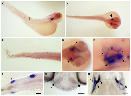
Dr-S100S expression pattern by whole mount in situ hybridization. Five day old zebrafish larvae were hybridized with RNA antisense probe. Panels A) to E), whole mounts; panels F) to H), sectioned after hybridization. A) Lateral view, expression in neuromasts (arrows) and otic placode is visible. B) Dorsal view, staining in both otic placodes (arrowhead) is seen. C) Magnification, neuromasts in trunk and tail are labeled. D) Frontal view of the head region, expression in neuromasts (arrow) and the otic placode (arrowhead) can be seen. E) Magnification of the ear region, expression in several spots (arrowheads) in the otic placode and in climbing fibers (arrows). F) Expression in tail neuromasts, all hair cells seem to be stained (arrow), the ring of hair cell nuclei is devoid of staining. Scale bar 10 μm. G) Expression in a symmetrical pair of neuromasts in the head region, scale bar 30 μm. H) Staining in the ear, anterior is up. Hair cells underlying the otoliths show strong expression (arrowheads). A neuromast (arrow) is also visible on each side. Scale bar 30 μm.
|
