|
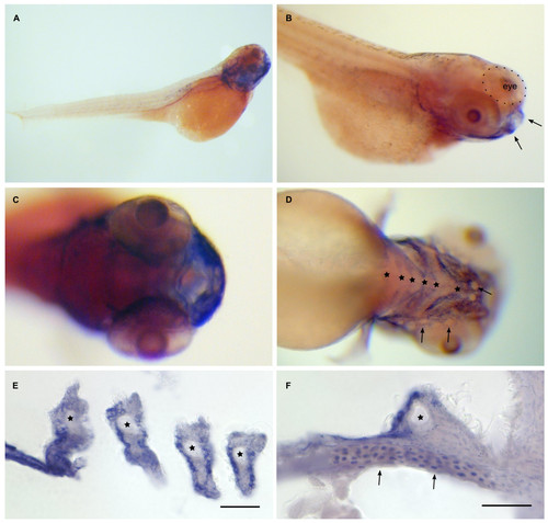
Dr-S100A10b expression pattern by whole mount in situ hybridization. Five day old zebrafish larvae were hybridized with RNA antisense probe. Panels A) to D), whole mounts; panels E) to F), sectioned after hybridization. Scale bars, 30 μm. A) Lateral view shows expression in the lower jaw. B) Lateral oblique view, lip region (arrows) expresses S100A10b. C) Frontal view (ventral to the right) shows expression in the mouth region. D) Ventral view (anterior is to the right), expression is visible in six branchial arches (asterisks), neuromasts (arrows) are not stained. E) Expression in branchial arches (asterisks) is limited to the epithelial layer. F) The seventh branchial arch (asterisk) is also expressing A10b, as well as cells of the pectoral fin (arrows).
|

