
Fig. 5
- ID
- ZDB-FIG-080325-65
- Publication
- Wilkins et al., 2008 - Mtx2 directs zebrafish morphogenetic movements during epiboly by regulating microfilament formation
- Other Figures
- All Figure Page
- Back to All Figure Page
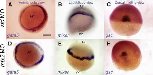
Normal endoderm and shield specification in mtx2 morphants. Whole-mount in situ hybridization for faust/gata5 (A, D) and mixer (B, E) shows endoderm specification in mtx2 MO injected embryos (D, E). The blastoderm margin appears a little irregular in mtx2 MO injected embryos (D–F). Goosecoid (gsc) is expressed in the shield of mtx2 MO injected embryos (F) but is reduced when compared with std MO injected embryos (C). All embryos are at shield stage. Panels A and D are animal pole views; panels B and E are lateral or oblique views; panels C and F are dorsal midline views. AP = animal pole, VP = vegetal pole. Scale bar = 200 μm in panel A (applies to panels A–F). |
| Genes: | |
|---|---|
| Fish: | |
| Knockdown Reagent: | |
| Anatomical Terms: | |
| Stage: | Shield |
| Fish: | |
|---|---|
| Knockdown Reagent: | |
| Observed In: | |
| Stage: | Shield |
Reprinted from Developmental Biology, 314(1), Wilkins, S.J., Yoong, S., Verkade, H., Mizoguchi, T., Plowman, S.J., Hancock, J.F., Kikuchi, Y., Heath, J.K., and Perkins, A.C., Mtx2 directs zebrafish morphogenetic movements during epiboly by regulating microfilament formation, 12-22, Copyright (2008) with permission from Elsevier. Full text @ Dev. Biol.