
Fig. S1
- ID
- ZDB-FIG-070914-75
- Publication
- Daggett et al., 2007 - Control of morphogenetic cell movements in the early zebrafish myotome
- Other Figures
- All Figure Page
- Back to All Figure Page
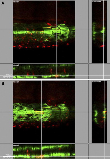
Cap1-deficient adaxial cells can adopt the slow-twitch myosin-expressing (F59-positive) fate and migrate to appropriate final positions. Shown are three-dimensional reconstructions of confocal images of slow-twitch, F59-positive (green) and Cap1-deficient, rhodamine-dextran-labeled transplanted (red) trunk muscle cells at the 26-somite stage. F59-expressing transplanted cells are yellow. Lateral, dorsal and transverse views of confocal sections containing transplanted cells are shown. Crosshairs highlight the position of the same cell in each view. Anterior is to the right in lateral and dorsal panels. F59-positive, Cap1-deficient transplanted cells can be found in normal slow muscle fiber positions, demonstrating that although Cap1 is required for normal pre-migratory adaxial cell behaviors, it is not required for slow muscle cell fate or for position in the postmigratory, superficial slow fiber (SSF) layer (A) and the non-migratory muscle pioneer (MP) cluster (B) in mosaic embryos. |
Reprinted from Developmental Biology, 309(2), Daggett, D.F., Domingo, C.R., Currie, P.D., and Amacher, S.L., Control of morphogenetic cell movements in the early zebrafish myotome, 169-179, Copyright (2007) with permission from Elsevier. Full text @ Dev. Biol.