
Fig. 2
- ID
- ZDB-FIG-070914-70
- Publication
- Daggett et al., 2007 - Control of morphogenetic cell movements in the early zebrafish myotome
- Other Figures
- All Figure Page
- Back to All Figure Page
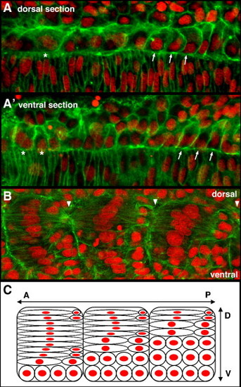
Adaxial cell behaviors occur with a dorsal to ventral progression within the developing myotome. Anterior to left in all panels. (A) In a more dorsal plane of adaxial cells adjacent to the notochord, the more anterior cells are broadening (*) while more posterior cells are undergoing interleafing behaviors (arrows). (A2) In a slightly more ventral plane, the anterior adaxial cells are beginning to lose epithelial shape and to accumulate cortical actin, characteristic of the early interleafing stage (*), while the more posterior adaxial cells still predominantly display a pseudo-epithelial appearance (arrows). (B) A lateral image of 3 adjacent somites, focused just lateral to the notochord surface, shows that, in each somite, the more dorsal adaxial cells in the somite have undergone further elongation and intercalation than those more ventral. Arrowheads highlight somite boundaries. (C) Schematic representation of panel B, illustrating both that the majority of more dorsal cells within the myotome are further advanced in their behaviors than the more ventral, and further, that within a given dorsoventral plane, the adaxial cells in more anterior somites are more advanced in their behaviors than those in more posterior somites as is apparent in Fig. 1. A, anterior; P, posterior; D, dorsal; V, ventral. |
Reprinted from Developmental Biology, 309(2), Daggett, D.F., Domingo, C.R., Currie, P.D., and Amacher, S.L., Control of morphogenetic cell movements in the early zebrafish myotome, 169-179, Copyright (2007) with permission from Elsevier. Full text @ Dev. Biol.