
Fig. 4
- ID
- ZDB-FIG-070620-42
- Publication
- Hans et al., 2007 - Changes in retinoic acid signaling alter otic patterning
- Other Figures
- (all 9)
- All Figure Page
- Back to All Figure Page
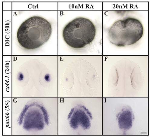
Increased RA signaling compromises lens specification. (A-F) Assessed both by morphology at 50 hpf (DIC microscopy, A-C) and cx44.1 expression at 24 hpf (D-F), the lens is reduced in size or completely lost after the application of 10 or 20 nM RA, compared with wild-type embryos. (G-I) Compromised lens specification is already evident at preplacodal stages (five-somite stage, 5S), as indicated by pax6b labeling. (A-C) Lateral views of live eyes with anterior to the left and dorsal towards the top. (D-I) Dorsal views with anterior towards the top. Scale bar: 30 µm for A-C; 50 µm for D-F; 35 µm for G-I. |
| Genes: | |
|---|---|
| Fish: | |
| Condition: | |
| Anatomical Terms: | |
| Stage Range: | 5-9 somites to Prim-5 |