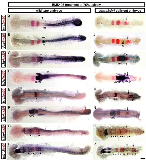
Development of supernumerary rhombomeres and their mirror-image patterning in cdx1a/cdx4-deficient zebrafish embryos with compromised RA signaling. Changes in rfng, foxb1.2 (mar), hox (purple) and krx20 (red) gene expression visualized in wild-type (A-H) and cdx1a/cdx4-deficient (I-P) embryos treated with the retinoic acid (RA) receptor inhibitor BMS493 (BMS) at mid-gastrulation (75% epiboly, 8 hpf). (A,I) hoxc4a expression in r8 is lost in wild-type (bracket with asterisk in A) and cdx1a/cdx4-deficient (I) embryos with compromised RA signaling. (B,J) RA inhibition reduced expression of the r7/8 marker hoxd4a in wild-type (bracket in B) and cdx1a/cdx4-deficient (J) embryos. (C,K) hoxd3a expression in the hindbrain posterior to r5 is reduced in wild-type embryos (bracket in C) and is limited to a central domain of the CNS in cdx1a/cdx4-deficient embryos (K). (D,L) hoxb3a, which is normally expressed posterior to the r4/r5 boundary (bracket in D), is expressed in a central domain in the CNS of cdx1a/cdx4-deficient embryos that includes the krx20 r5 and ectopic expression domains (L). (E,M) hoxb1a is expressed in r4 of wild-type (E) and cdx1a/cdx4-deficient (M) embryos. The latter also shows an additional hoxb1a-positive domain of expression in the posterior CNS. (F,N) hoxa2b is strongly expressed in r2 and r3 and weakly in r4 in wild-type (F) and cdx1a/cdx4-deficient (N) BMS-treated embryos. (G,O) rfng is expressed in six and nine boundary-like stripes (arrowheads) in wild-type (G) and cdx1a/cdx4-deficient (N) BMS-treated embryos, respectively. (H,P) foxb1.2 is expressed in nine boundary-like stripes (arrowheads) in cdx1a/cdx4-deficient BMS-treated embryos (P), compared with the six stripes seen in their wild-type siblings (H). For each condition, a minimum of 44 embryos from at least three independent experiments was analyzed at the equivalent of the 20-somite stage. More than 82% of embryos displayed the phenotype shown. Representative embryos were dorsal flat-mounted, anterior to the left. Ectopic rhombomere-like krx20 domain of expression is labeled with an arrow. Position of somite 3, the hindbrain-spinal cord transition in wild-type embryos, is indicated with a white arrowhead. Supernumerary rhombomeres are labeled r'. Scale bar: 100 µm.
|

