
Fig. 1
- ID
- ZDB-FIG-061212-1
- Publication
- Wang et al., 2006 - Myocyte-specific enhancer factor 2A is essential for zebrafish posterior somite development
- Other Figures
- All Figure Page
- Back to All Figure Page
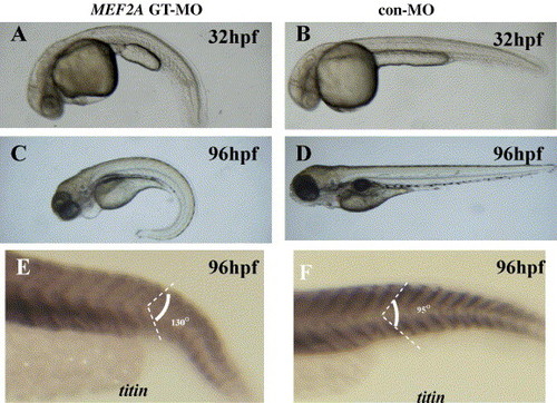
MEF2A morphants display a downward tail curvature and have U-shaped posterior somites although the somite borders are generated normally. (A–F) Lateral views with anterior to the left. The overall morphology of a MEF2A MO-injected (A and C) and control MO-injected (B and D) embryos at 32 hpf (A and B) and 96 hpf (C and D). Note that the MEF2A morphants display a downward tail curvature (A and C) (n = 50 embryos). (E and F) Lateral views of titin expression, which marks each segment border in MEF2A GT-MO-injected (E) and control MO-injected (F) embryos at 96 hpf. Injection of control MO results in chevron-shaped somites with an angle of 95° (n = 20 embryos), while injection of MEF2A GT-MO results in a more obtuse angle of the somite (130°) (n = 19 embryos). |
| Gene: | |
|---|---|
| Fish: | |
| Knockdown Reagent: | |
| Anatomical Term: | |
| Stage: | Day 4 |
| Fish: | |
|---|---|
| Knockdown Reagent: | |
| Observed In: | |
| Stage Range: | Prim-15 to Day 4 |
Reprinted from Mechanisms of Development, 123(10), Wang, Y., Qian, L., Dong, Y., Jiang, Q., Gui, Y., Zhong, T.P., and Song, H., Myocyte-specific enhancer factor 2A is essential for zebrafish posterior somite development, 783-791, Copyright (2006) with permission from Elsevier. Full text @ Mech. Dev.