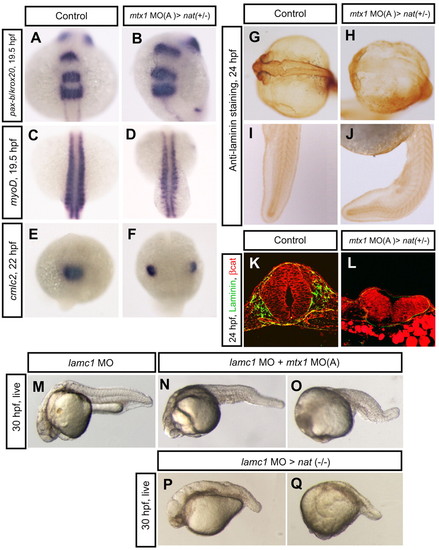
Regional specification and ECM formation in mtx1 MO-injected heterozygous natter embryos. (A,B) Dorsal views of hindbrain, (C,D) somite and (E,F) heart regions. pax-b and krox20 expression in the midbrain-hindbrain boundary and rhombomeres 3 and 5 at 19.5 hpf (A,B), and myod expression in the segmented somites and adaxial cells at 19.5 hpf (C,D), appeared unaffected in mtx1 MO-injected heterozygous natter embryos. (E,F) cmlc2 expression in the myocardial cells at 22 hpf reveals that mtx1 MO-injected heterozygous natter embryos displayed cardia bifida. (G-L) Laminin immunostaining at 24 hpf. Laminin deposition appeared to be greatly reduced in the hindbrain region of mtx1 MO-injected natter heterozygous embryos (H), but not in the trunk and tail (J). (K,L) Transverse sections of embryos (anterior region) immunostained for β-catenin (red) and Laminin (green). Laminin deposition was greatly downregulated and the head structure collapsed in mtx1 MO-injected heterozygous natter embryos (L). (M-Q) Lateral views of bright field images at 30 hpf, anterior to the left. Embryos injected with laminin c1 MO at the 1-cell stage exhibited shortened body axis and defects in notochord differentiation (M). Embryos injected with lamimin c1 MO at the 1-cell stage and mtx1 MO(A) into the YSL at the 1000-cell stage showed enhanced phenotypes in the hindbrain region (N) or entire head region (O). Homozygous natter mutant embryos injected with laminin c1 MO (P,Q).
|
