|
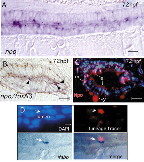
Expression and function of npo in the endoderm. (A) Mid-sagittal section of 72 hpf embryo labeled for npo by in situ hybridization, showing punctate, heterogeneous staining in epithelial cells. (B) Double in situ hybridization to npo (dark blue; arrowheads) and foxa3 (brown), showing npo expression in a subset of endoderm-derived epithelial cells. (C) Anti-Npo immunofluorescence (red) at 72 hpf shows expression primarily in gut epithelium, and also in scattered subepithelial mesenchymal cells. The yolk signal is caused by autofluorescence. (D) Mosaic analysis: wild-type cell (arrow) in npo-/- host shows cell autonomous rescue of ifabp expression. Panels show nuclei (DAPI; blue), lineage tracer (rhodamine dextran; red) and labeling for ifabp (dark blue). e, epithelium; m, mesenchyme; y, yolk. Scale bars: A,B, 10 μm; C, 15 μm; D, 7 μm.
|