
Fig. 6
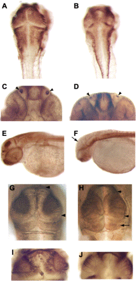
col mutant embryos display a reduction of neural tissue and regional posteriorization of the neuroectoderm. (A,B) Dorsal views of wild-type (A) and col mutant (B) embryos at 2 dpf labeled with zn5 antibody. The developing brain in col mutants appears laterally reduced (B). (C,D) Ventral views of the heads of wild-type (C) and col mutant (D) embryos at 2 dpf labeled with anti-acetylated tubulin antibody with arrowheads indicating the olfactory organs that are reduced in size in col mutants. (E,F) Lateral views of wild type (E) and col mutants (F) labeled with anti-acetylated tubulin antibody at 2 dpf. Arrow in (E) marks staining in the tectum of wild-type embryos. A dramatic reduction in staining in seen in col mutants (F). (G,H) Dorsal views of wild-type (G) and col mutant (H) embryos at 48 hpf labeled with zn12 antibody with arrowheads marking the anterior–posterior extent of the tectum. The size of the tectum is reduced in col mutants whereas the cerebellum is larger (arrow in H). (I,J) A drastic reduction in tectal projections is evident in 2 dpf col mutants (J) labeled with zn5 antibody as compared to wild type (I). |
Reprinted from Developmental Biology, 267(1), Nambiar, R.M., and Henion, P.D., Sequential antagonism of early and late Wnt-signaling by zebrafish colgate promotes dorsal and anterior fates, 165-80, Copyright (2004) with permission from Elsevier. Full text @ Dev. Biol.